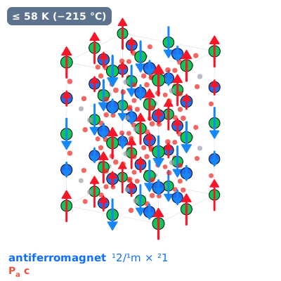
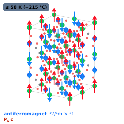
No net magnetisation, and up and down sublattices are related by a shift or by inversion, so the bands stay spin-degenerate: a conventional antiferromagnet.
Spin group ¹2/¹m × ²1: each symmetry marked ¹ keeps every spin, ² swaps up and down.
ordered ≤ 58 K (−215 °C)Colour: where that sits relative to 25 °C. Saturation: how much margin it has.
Structure · 10 properties
Magnetic group
Pa c BNS 7.27
Parent space group
P 21/n No. 14
Spins
collinear · net moment 0 μB per cell
Propagation
[1/2 0 1/2] 1k
Cell
a 11.2191 · b 5.5703 · c 15.7428 Å · α 90° · β 89.96° · γ 90°
Transition
58 K (−215 °C)
Measured at
1.5 K
Magnetic ions
Ni, Os
Largest moment component
1.9 μB
Method
NPD
Symmetry
Stereogram of the magnetic point group: each operation applied to one direction. Filled = upper hemisphere, ring = lower; red operations include time reversal. Lines are mirrors; polygons are 2/3/4/6-fold axes; the centre ring is inversion.
sha256 of this copy: 56a76c2ca8fb13fe4d7368813324c9a99979b14863d0b5fb8a238f359a01d6fb The mirror keeps the bytes Bilbao served, so the original should hash the same unless MAGNDATA has revised the entry since: shasum -a 256 1.592_Pb2NiOsO6.mcif.